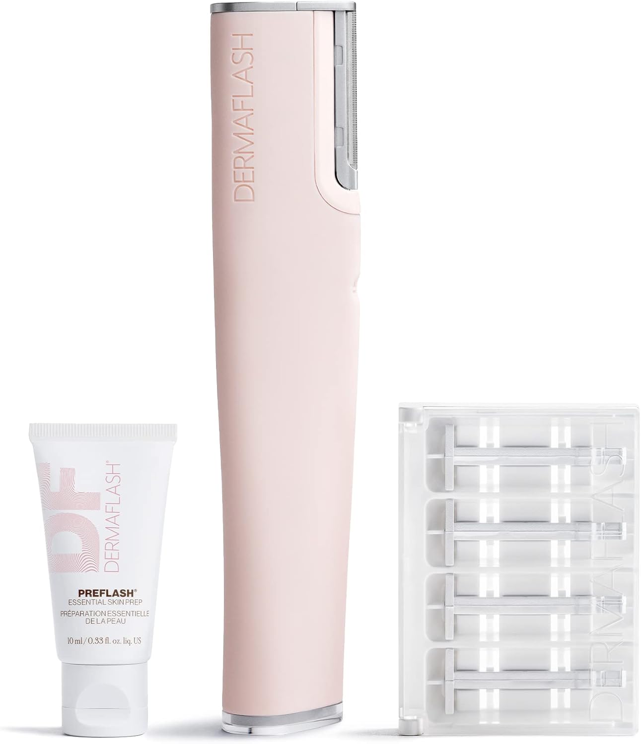
DERMAFLASH LUXE+ Device, Anti,Aging, Exfoliation, Hair Removal, and Dermaplaning Tool with Sonic Edge Technology and 4 Weeks of Treatment
FREE Shipping
DERMAFLASH LUXE+ Device, Anti,Aging, Exfoliation, Hair Removal, and Dermaplaning Tool with Sonic Edge Technology and 4 Weeks of Treatment
- Brand: Unbranded

Description
For most people, this will probably be an effective product, but it’s always important to keep a few tips in mind when using the device: By and large, most customers have been happy with DermaFlash, but there were a few who said it didn’t live up to the advertising. is definitely an exception. Glowy skin sans makeup has always been my aesthetic, so when something promises to nix peach fuzz, I’m game to try at least once. Admittedly, I was both intrigued and skeptical walking into this because, well, shaving one’s face isn’t something you do every day. Nonetheless, it’s an adventure well worth taking.
A: Step 1: PREFLASH(TM): The essential first step, wash your face with the cleanser provided, rinse, and towel your face dry. Place a small amount of PREFLASH onto wet hands and create lather. Massage onto face, rinse well, and pat dry. The majority of users have been very happy with this product. They found it to be very gentle on their face. It removed the dead skin and peach fuzz on the face and left it glowing and clean-looking.
More From Feelunique
Others mentioned glitches like a charging port that stops working after only a few uses, or the blade eject mechanism failing. A: This product does work for most customers. The majority of users have been very happy with the results they’ve gotten after even one usage. It makes the skin smoother and refreshes it. Some users have developed pimples or a rash, but most have not. Buzzing for something new and exciting? Taking your skin care routine to a whole new glowing level with the DERMAFLASH LUXE+ 2.0 and enjoy pro-level dermaoplanning from the comfort of your bathroom with the widely acclaimed exfoliating-meets-shaving tool. A: If you have any cuts or abrasions on your face, you can’t use the device. In addition, if you have eczema or any other skin condition, you should avoid using this product.
Customers said that following use their face felt invigorated. They found it to be worth the money, especially since laser treatments that would accomplish hair removal are much more costly. Now that we’ve answered up your biggest question, it’s time to dive into the benefits. What is dermaplaning? Some users did say they got redness or a rash from the product, and even ingrown hairs and pimples in a couple of cases. It’s a good idea to try it out on a small area of skin first. If you have any kind of skin condition, including bad acne, you should avoid using this product. Go to your doctor, or to a dermatologist, before beginning to use DermaFlash. It’s important to make sure that it’s the right product for you. Is There Anything Else You Should Know About DermaFlash? Note that some of the products can be used on the entire body, making them more valuable and time and money-saving than DermaFlash. Is DermaFlash Safe? – Warnings & ComplaintsDermaFlash 2.0 Luxe– The DermaFlash 2.0 Luxe allows for 8 treatments, and the kit includes a 20 ml PREFLASH Cleanser, 20 ml POSTFLASH Moisturizer, Travel Edge Tray with two full-size edges, a travel bag, and a DERMAPROTECT Daily Defense Broad Spectrum SPF 50+. (4) Step 2: DERMAFLASH(R): With a new Edge loaded, take the device and turn it on with the power button. It will begin to gently vibrate. If you press it again, it will vibrate more intensely. Pressing it a third time turns it off. Start near your ear and hold your skin taut while you use the device. Hold your DERMAFLASH(R) LUXE at a 45o angle. Run the device over your face gently using short, feathery strokes. Do not get it near the surface of your eyelids, lips, or nose. There were some users that said the treatment made their face red afterward, led to a rash or pimples. A couple of users said that it actually did create stubble or ingrown hairs when the peach fuzz grew in – contrary to what the company advertises. In most cases, it removes the outer layer of skin and peach fuzz from the face leaving it smooth and replenished. In some cases, however, it causes skin issues like pimples or a rash. Purified Water, Sodium Lauroyl Methyl Isethionate, Acrylates Crosspolymer-4, Cetyl Hydroxyethylcellulose, Papain, Phospholipids, Glucosamine HCI, Aphanizomenon Flos-Aquae Powder, Salix Alba (Willow) Bark Extract, Salicylic Acid, Yeast Extract, Glycerin, Leuconostoc/Radish Root Ferment Filtrate, Phenoxyethanol, Urea, Potassium Sorbate, Sodium Benzoate, Sodium Hydroxide, Disodium EDTA Moisturizer
DermaFlash is a product that seems to be great for a lot of people and not so good for some others. DermaFlash – LUXE Anti-Aging Dermaplaning Exfoliating Facial Treatment – Health Insiders Images DermaFlash Ingredients Cleanser
- Fruugo ID: 258392218-563234582
- EAN: 764486781913
-
Sold by: Fruugo